What is cancer?
Cancer is not a single illness: there are very many different types. Some cancers may stay almost unchanged for several years and have no impact on life expectancy. At the other end of the spectrum there are rare cancers which may prove fatal quite shortly after they have been discovered. In the same way that the term ‘infection’ embraces illnesses as far apart as the common cold, a boil, malaria and tuberculosis, the spectrum of malignant disease is almost equally varied, in both behaviour and seriousness although, of course, cancer is not contagious.
Loss of control
A lump of human tissue the size of a sugar cube contains roughly a thousand million cells. These are the minute building blocks from which our bodies are made, visible only down the microscope. It is quite amazing that the billions of cells in a human body normally function in perfect harmony, every cell knowing its place and doing the job that it was designed to do. Most cells have a finite lifespan: millions of new ones are produced every day to replace those lost through old age or wear and tear.
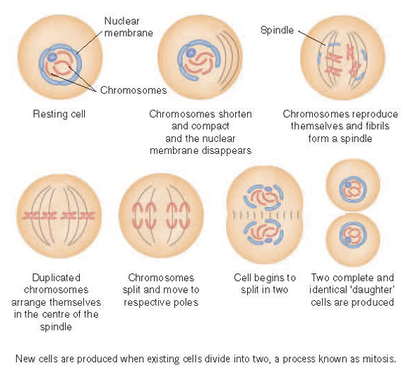
New cells are produced when existing cells divide into two, a process known as ‘mitosis’ Except in children and younger people who are growing, there is normally a perfect balance between the numbers of the cells that are dying and those that are dividing. Normally exactly the right amounts of new cells are produced to replace those that are being lost. The control mechanisms involved are exceedingly complex. Loss of control can lead to an excess of cells, resulting in a tumour.
However, it is important to realise that only a small minority of tumours are cancerous. Most tumours are localised accumulations of normal or fairly normal cells and are benign. A wart is a common example.
The development of a cancer involves a change in the quality of the cells as well as an increase in quantity: they change in both appearance and behaviour. They become more aggressive, destructive and independent of normal cells. They acquire the ability to infiltrate and invade the surrounding tissues. In some instances the cells may also invade lymphatic and blood vessels and thus spread away from the ‘primary’ growth to other places. In time these cells may cause the development of secondary growths, known as ‘metastases’, in the lymph glands and other organs such as lungs, liver and bones.
Genes
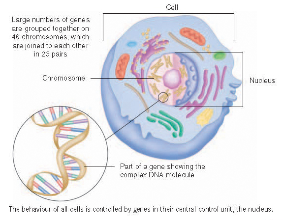
The behaviour of all cells is controlled by genes in their central control unit, the nucleus. Each cell nucleus contains approximately 20 – 25 thousand genes. The genes are minute, highly concentrated packets of information and instructions stored in coded form in a complex chemical molecule known as ‘DNA’. Large numbers of genes are grouped together in strands looking rather like short pieces of string, which are just visible under the microscope. These are the chromosomes, which are joined to each other in pairs, 23 pairs in total.
A human being develops in the uterus from a single cell. This first cell is formed by the fertilisation of an ovum (egg) produced in one of the mother’s ovaries by a sperm produced in one of the father’s testes. It divides to produce two ‘daughter’ cells and then they divide, resulting in four cells. Successive divisions lead to rapid growth. Mitosis involves replication of all the genetic information so that all the cells in the developing microscopic organism or ’embryo’ each have their own full complement. This process continues as the embryo develops into a ‘fetus’ and then eventually into a newborn baby.
The genetic information contained within the first cell is what determines the physical characteristics of the whole human being that ultimately develops from it and the total amount of genetic information is often referred to as that person’s ‘genome’. However, once the body is fully formed, most of this genetic information is no longer required by any particular individual cell. All it needs is the information required to enable it to perform its own designated role. Instructions on how to perform other roles are redundant. The important pieces of information that are ‘switched on’ in particular cells govern the characteristics and behaviour of those cells and the properties of the particular tissues that they constitute.
Cancer genes
There are particular genes known as ‘oncogenes’ which are present in normal cells, where they may either be dormant or play a part in controlling cell behaviour and division. DNA damage caused, for example, by tobacco smoke, ultra-violet light or certain viruses can trigger faults or ‘mutations’ in these genes, resulting in increased and abnormal activity of the gene. This can cause the cell to behave in an antisocial way and eventually to become malignant (cancerous).
In addition to oncogenes, each cell contains ‘tumour suppressor genes’ whose normal job is to restrain it from dividing. Many cancers are caused by damage which reduces the activity of a tumour suppressor gene.
Genes are crucial not only to the development of malignancy, but also to the subsequent behaviour of a cancer and its response to treatment. For example, some genes are responsible for the manufacture of proteins important for a cancer’s ability to invade adjacent tissues and to spread to distant parts of the body, triggering the development of metastases. Other genes can cause the cell to produce self-stimulating or blood vessel supply increasing ‘growth factors’, or to eliminate anti-cancer drugs. Even the process of cell death is under genetic control. Genetic damage may result in cells failing to die, which can be an important factor both in the development of a cancer and in its resistance to treatment with radiotherapy or drugs.
The development of a cancer involves an accumulation of successive genetic abnormalities over some years both before and after the cell starts behaving in a malignant fashion. Further gene mutations after the cancer has started can result in some of the cancerous cells behaving differently from others. This may cause the growth to ‘change its spots’ at some stage. The behaviour of the cancer and the long-term result of treatment depend ultimately on those cells that have the most anti-social characteristics and on those that are best able to resist treatment aimed at destroying them.
Speed of growth
Most cells divide at the most every few days, some much more slowly. Given that almost all cancers start off as a result of a genetic abnormality in a single cell, and that there are about a thousand million cells in a sugar cube-sized lump, it follows that most cancers begin a very long time before they become apparent. Most cancers are rather larger than the size of a sugar lump when they are discovered and many will by then have been present, growing slowly, for 10 to 20 years. There is, however, a large variation in the time a tumour takes to double in size. This ‘doubling time’ can vary from a few days to very many years, and indeed many cancers are effectively dormant and would remain undiscovered unless detected by screening, which is discussed later. However, for most common ‘active’ cancers the average doubling time is of the order of two to three months.
An important factor in the rate of growth is the blood supply to the cancer, from which it gets its nourishment. The triggering action of growth factors on both the formation of new blood vessels and cancer cell division depends on these molecules linking up with ‘receptor’ molecules on the surface of the cells. Drugs that inactivate or ‘block’ these growth factor receptors now play an important rôle in controlling some types of cancer.
Effects of cancer
It can sometimes be difficult to understand just how an excessive number of abnormal cells can in certain circumstances be a threat to life. The serious effects of malignant disease occur as a result of progressive infiltration and destruction of the surrounding normal tissues and/or other parts of the body to which the cancer has spread, such as the liver, bones or lungs, disrupting their normal function. It is fairly unusual for localised cancers to be fatal. Most of those who die from cancer have widespread or ‘metastatic’ disease. However, in addition to these ‘physical’ processes, cancers can cause progressive debilitation by producing a wide range of toxic chemicals acting both locally and, via the bloodstream, throughout the body. These chemicals can be responsible for symptoms such as loss of weight and weakness.
Classification
Most cancers are graded according to the extent to which the cells are different from normal. In well-differentiated (sometimes called ‘low grade’ or ‘grade 1’) cancers some of the normal cellular architecture is maintained and the cells do not seem to be dividing frequently. Some may still be retaining some ability to perform their original specific tasks. At the other end of the spectrum are poorly differentiated (‘high grade’ or ‘grade 3’) cancers in which the cells have changed so much that they are now very different from normal cells and have completely lost their ability to perform their appointed tasks. These cancers tend to be faster growing and more aggressive, and to carry a less favourable prognosis. Moderately differentiated cancers are in between. Prostate cancers have their own grading ‘Gleason score’ system and most are graded from 6 to 8, where 6 is regarded as low grade and 8 as high grade.
Cancers are classified according to the type of normal cell from which they originated, not according to the tissues into which they may have spread. This is what might be called the primary classification. Cancers in each category will be graded as described above, and their growth and extent of spread will also be assessed in a process known as ‘staging’, to be discussed shortly. As far as primary classification is concerned, almost all cancers can be placed in one of the following groups.
Carcinomas
These are by far the most common types of cancer. They originate from cells lining body surfaces, including the skin and a wide variety of internal linings. Among these are those of the mouth, throat, bronchi (the tubes carrying air in and out of the lungs), oesophagus (the swallowing tube or gullet), stomach, bowel, bladder, uterus (womb) and ovaries, and the linings of ducts in the breasts, prostate gland and pancreas.
There are different types of carcinomas named according to the appearance of the normal cells from which they arose. ‘Squamous carcinomas’ arise particularly in the skin, lung, mouth, throat and oesophagus; ‘adenocarcinomas’ arise particularly in the breast, bowel, lower oesophagus, stomach and ovary; ‘transitional cell carcinomas’ arise chiefly in the bladder and ‘small cell carcinomas’ also occur in the lung.
Sarcomas
These arise from supportive rather than surface lining tissues, such as those of bone, fat, muscle and the strengthening ‘fibrous tissue’ found in most parts of the body.
Lymphomas
These originate from cells known as ‘lymphocytes’ which are found throughout the body, particularly in the lymph glands and blood. They are a very important component of the body’s immune system. Lymphomas are divided into ‘Hodgkin’s disease’ and the ‘non-Hodgkin’s lymphomas’, according to the cell type affected.
Leukaemias
These arise from the cells in the bone marrow which make the white blood cells. The white blood cells or ‘leukocytes’ are crucial to the body’s defence system against infection. In leukaemia there is a greatly increased concentration of abnormal white cells in the blood, which causes problems both because the abnormal cells often don’t function properly and because they restrict the space within the bone marrow for new normal blood cells to be made.
Myeloma
This is a malignancy of the ‘plasma cells’ in the bone marrow which produce antibodies – the proteins that help to fight infections.
Germ cell tumours
These develop from those cells in the testes and ovaries responsible for the production of ova and sperm. They include ‘teratomas’ and ‘seminomas’.
Melanoma
This type of skin cancer arises from the skin pigment-producing cells or ‘melanocytes’.
Gliomas
These develop from cells of the supportive tissue of the brain or spinal cord.
Pre-cancers
Finally, it is important to mention the fairly common potentially pre-cancerous change which is quite often picked up in apparently healthy people who undergo ‘screening’ tests such as cervical smears and mammograms (breast X-rays – see next chapter). It particularly affects the inner surfaces of the cervix (neck of the womb) and breast ducts and it can also be found in the bladder, prostate, bowel and skin. Such change is referred to as ‘carcinoma in situ’ or ‘intraepithelial neoplasia’. This means that the cells on the very surface have a malignant appearance when seen though a microscope, but show no sign of having begun to behave malignantly by invading any of the tissue immediately beneath the surface lining.
Carcinoma in situ has no ability to spread via the lymphatic or blood systems and in itself poses absolutely no threat to life. It does, however, carry a very variable risk of eventually becoming truly cancerous if left untreated.
KEY POINTS
-
Cancers start off as a consequence of gene damage in a single cell and usually take many years to become apparent
-
Cancers vary enormously in appearance, behaviour and prognosis (outlook)


